Introduction to Eye Tracking: The Anatomy of the Eye
 The eye is a complex organ consisting of various muscles, tissues, and nerve sensors, which work together to create the phenomenon we know as vision. The ability to see, for those of us who possess the gift of sight, is something we take for granted. It seems simple enough– you open your eyes and voila! You see the world around you; however, it isn’t a magic trick. We write about eyes and vision in just about every article on Eye Tracking Update, but to get a better idea of how eye tracking works, you should understand the biology behind this seemingly effortless ability.
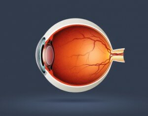
To begin, here are some basic descriptions of the key parts of the anatomy of the eye:
Sclera: The white portion of the eye, which is a dense tissue containing blood vessels and providing a surface for attaching the external muscles of the eye.
Pupil: The round, black opening in the center of the iris that allows light to pass through the eye and onto the retina.
Iris: The colored part of the outer eye. This thin muscle constricts or dilates to adjust the diameter of the pupil, thus controlling the amount of light that enters the eye.
Cornea: The transparent tissue that covers the front of the eye including the pupil and iris. It refracts light as it enters the eye and is responsible for most of the eye’s focusing abilities.
Lens: After light enters through the pupil, it passes through the lens, which focuses the light and projects it onto the surface of the retina in the back of the eye.
Retina: A light sensitive tissue that lines the inner surface of the back of the eye. It transforms light that enters through the pupil and passes through the lens into nerve signals that are converted into an image by the visual cortex of the brain.
All of these components function simultaneously to allow us to see our surroundings. The resulting picture, called the visual field, is a combination of two primary types of vision with distinct functions and characteristics. The most focused, yet smallest portion of our visual field, is called foveal vision. Objects within the scope of foveal vision are clear and colorful. The majority of the scene we see, however, falls in our peripheral vision, which is blurry and less colorful. It allows us to detect movements and color and shape contrasts that warrant further examination by the foveal vision; peripheral vision also permits vision under low light conditions.
These two different types of vision are a result of the two kinds of light receptor cells found in the retina: the cone cells and the rod cells. Foveal vision is created by cone cells that are tightly packed in a small area in the center of the retina and only account for 6% of the total retinal light receptors. Cone cells require the most light for creating a clear, detailed image. The rod cells account for the other 94% of light receptors in the retina. They require less light but, as mentioned above, create the blurry, less colorful qualities of peripheral vision.
An Introduction to Eye Tracking : Part 1 – How does the eye work?
Related articles:
- Eye Tracking: Challenging the Biological Monopoly
- Where’s Waldo?… Eye Tracking Only Registers Half of the Visual Search Process
- How Does Eye Tracking Detect the Object of Visual Attention?
- Fixations and Saccades in Eye Tracking
- Eye Tracking … Baby-Style
- Pupil Tracking in Airport Security: Can Body Language Indicate Terrorist Intent?
- Eye Tracking History: An Early Eye Tracking Apparatus
- Eye Controlled Video Games? Better Late Than Never
- Eye Tracking on the Cheap: Making MacGyver Proud
- Neuromarketing Eye Tracking Helps Campbell’s Soup Get a Makeover

